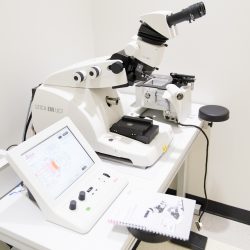
The Evident imaging equipment is part of The University of Texas at Dallas / Evident Discovery Center. This center is a collaborative effort between UTD and Evident Scientific which includes the core facility, on-site training of Evident staff, hands-on microscopy workshops, and regular beta testing of new Evident imaging equipment.
Evident Scientific and Zeiss Imaging Instruments
The Evident Scientific imaging equipment is part of The University of Texas Dallas/Evident Scientific Imaging Reference Center. This center is a collaborative effort between The University of Texas Dallas and Evident Scientific establishing a state-of-the-art imaging core, including top-end Evident Scientific imaging facilities, hands-on training from Evident Scientific staff, an imaging summer school run cooperatively by Evident Scientific and UTD, and regular beta testing of new Evident Scientific imaging equipment.
-
Leica Stellaris STED Super Resolution Confocal Laser Scanning Microscope
Laser scanning confocal microscope equipped with a tunable white light laser system, stimulated emission depletion laser for super resolution microscopy, and tunable emission filters for fixed or live sample imaging in the visible range.
-
Evident Scientific FV4000 Confocal Laser Scanning Microscope
Laser scanning confocal microscope equipped with 10 lasers, 6 detectors, and tunable emission filters for fixed or live sample imaging in the visible and near-infrared range.
-
Evident Scientific VS200 Research Slide Scanner
210-Slide Scanning System for automated high-throughput imaging in brightfield, fluorescence, darkfield, and polarized light modes
-
Evident Scientific SpinSR Spinning Disk Super Resolution Microscope
Spinning Disk Confocal Microscope designed for high speed live cell imaging
-
Evident Scientific MPE-RS TWIN Multiphoton Microscope
Intravital Multiphoton Fluorescence Microscope with dual multiphoton laser system for simultaneous scanning and excitation
-
Evident Scientific cellTIRF-4Line System
Total Internal Reflectance Fluorescence system equipped with 4 laser lines with individual control
-
Zeiss EVO LS 15 SEM
Environmental Scanning Electron Microscope, Beam: 30 keV (W filament), High resolution (3nm)/high vacuum mode, Variable pressure mode (10-3000 Pa) for non-conductive specimen, Detectors: 3 SE, 1 BSD, and an Oxford Explore 30 EDS. Deben peltier cooling stage (-25° to +50°) for wet samples
-
JEOL JEM 1400+ TEM
Beam: 120 keV/LaB6, Resolution: Sub-nm, Camera: Gatan OneView (16 MPixel), Holders: Regular, High tilt, and Cryo. Tomography: SerialEM. Other associated tools: FEI Vitrobot plunger, Glow discharge grid cleaner, Gatan 665 pump station, and Leica series sample preparation tools in Histology Core.
Histology and Cell Culture Equipment

- Leica ASP300 S: Fully enclosed tissue processor
- Leica EM UC7: Ultramicrotome used for sample preparation for electron microscopy
- Leica RM2235: Manual microtome
- Leica CM1860: Clinical Cryostat
- Leica EM FC7: Cryochamber used for sample preparation for electron microscopy
- Leica ARCADIA C and H: Cold and hot paraffin embedding stations
- Leica EM ACE200: Carbon coater used for sample preparation for electron microscopy
- Leica EM KMR3 Glass Knifemaker
- NuAire Class II, Type A2 Biological Safety Cabinet
- NuAire Direct Heat In-Vitrocell Air-Jacketed CO2 Incubators
- 10X Genomic Xenium Analyzer: An imaging-based in-situ spatial gene expression platform providing subcellular resolution analysis of hundreds of RNA targets in the fresh frozen or FFPE tissue samples. You can select from pre-designed gene panels or create custom ones from 10X Genomics.
- 10X Genomic CytAssist: It simplifies the Visium workflow by enabling the rapid transfer of transcriptomic probes from tissues on standard microscope slides to Visium slides. Tissue preparation, staining and imaging occur on standard glass slides. After hybridization, the analytes are transferred to Visium slides for precise alignment within CytAssist. The remaining steps follow the standard Visium workflow outside of the instrument.


You must be logged in to post a comment.