This instrument has been decommissioned.
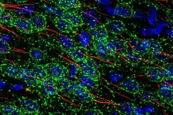
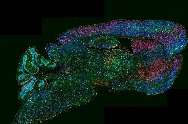
Laser Scanning Confocal Microscope equipped with 7 laser lines, a fully automated stage for whole tissue stitching, and live cell imaging capability
Location
Visuals


Capabilities
- Imaging diverse fluorophores in fixed or live samples
- Temperature, humidity, CO2 control
- Spectrally tunable detectors
- Ultra-fast resonant scanning (reduced photo-damage with live samples)
- 3D z-stacks, timelapse, multi-position timelapse
- FRAP/FLIP/Photoactivation, FRET (ratio or acceptor photobleaching)
Cost
Internal user $32/hour
Specs
Microscope
- Olympus IX83 fully motorized inverted microscope
- Zero Drift Compensator (IX3-ZDC2) laser-based autofocus
- Ultrasonic automated stage and multiple area time lapse stage control software, as well as well-plate navigator tool for multi-area, time-lapse, and multi-area mosaic image stitching
- Tokai hit stage top, live-cell incubation chamber
- Analog and digital in-out box for synchronization with external devices
- Facilitates integration of PicoQuant, FCS/FCCS, and FLIM hardware packages
Tandem Scanners
- Conventional scanner for high-definition imaging
- Resonant scanner for high-speed imaging (capable of 30 FPS@512×512 and 438 FPS@512×32)
Light Sources
- All Diode Lasers with 7 Laser Lines–405 nm, 445 nm, 488 nm, 514 nm, 561 nm, 594 nm, and 640 nm
- LED-based transmitted and epifluorescence illumination
Conventional Fluorescence Filters
- Dapi
- Green
- Red
Objectives
Air objectives:
- 25X; PLAN APO 1.25X, NA 0.04,WD 5.1 mm
- UPLSAPO10X2; U PLANS S-APO 10X, NA 0.4, WD 3.1 mm
- UPLSAPO20X; U PLAN S-APO 20X, NA 0.75, WD 0.65 mm
Silicone oil objectives:
- UPLSAPO30XS; UPLSAPO N 30X SI OIL, NA 1.05,WD 0.8MM, W/CC (with correction collar)
- UPLSAPO40XS; UPLSAPO N 40X SI OIL, NA 1.25, WD 0.3MM,W/CC
- UPLSAPO100XS; UPLSAPO 100X SI OIL, NA 1.35,WD 0.20MM,W/CC
Silicone oil provides better refractive index match to cells and tissue than water or traditional immersion oil for brighter and deeper cell/tissue imaging. Silicone Oil does not evaporate nor degrade permitting very long live cell/tissue time-lapse imaging with ease. Greater working distance than most traditional immersion objectives while still retaining high numerical aperture (30x – 1.05 NA 800 µm WD; 40x – 1.25 NA 300 µm WD; 100x – 1.35 NA 200 µm WD)
Plan Super Apochromat design fully compensates for both planar and chromatic aberrations from the UV to the near infrared region for precise multicolor colocalization imaging.
Dichroic(s)
- Conventional dichroic mirrors
Detectors
- 4 High-Sensitivity Peltier-Cooled GaAsP Spectral Confocal Detectors with Olympus TruSpectral High-Efficiency Volume Phase Hologram spectral detection system (1-100 nm adjustable emission bandwidth with 2 nm spectral resolution)
- Transmitted light detector for Brightfield/DIC Image Acquisition
Computer and Software
- HP Windows 7 64bit Confocal Workstation and 30” Monitor
- Complete Fluoview Acquisition & Analysis Software provides FRAP, FRET, Ratiometric Imaging Display & Analysis, 3D Image Acquisition & Display, Live 16 Channel Spectral Unmixing, Fluorescence Intensity Measurements, Time-Lapse Imaging, and More…
- Olympus cellSens Dimension provided for additional post-acquisition data analysis and processing
- Includes Olympus cellSens Count & Measure Solution for threshold-based object detection as well as automatic object measurement and classification
Includes Olympus cellSens 3D Deconvolution Solution utilizing Constrained Iterative Deconvolution algorithm for improved resolution, contrast and dynamic range.
Posted under Uncategorized


You must be logged in to post a comment.