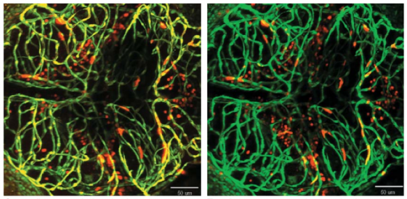
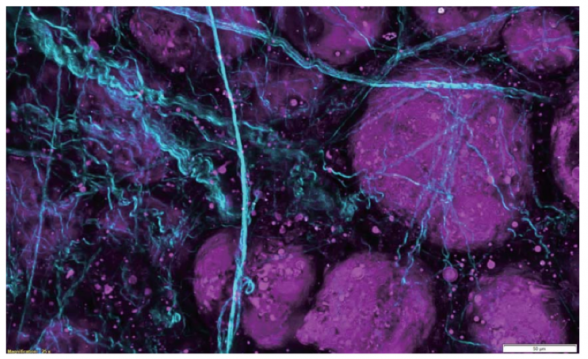
Intravital Multiphoton Fluorescence Microscope with dual multiphoton laser system for simultaneous scanning and excitation
Location
Visuals
Capabilities
- Multiphoton imaging of thick in vivo and in vitro samples
- Simultaneous photostimulation or dual-line imaging
- 405 nm UV photostimulation
- Long, working-distance objectives optimized for multiphoton
- High-speed extended IR (1300 nm) multiphoton imaging (30 FPS at 512 x 512, 438 FPS at 512 x 32)
- 4 non-descanned detectors (2 GaAsP and 2 multi-alkali PMTs) capable of conducting Fluorescence Lifetime Imaging Microscopy (FLIM)
Cost
Internal user $33/hour
Specs
Microscope
- Olympus IX83 fully motorized upright microscope
- Twin IR laser coupling optics with full laser enclosure for independent control of two IR lasers
- Motorized mirror assembly permits user-selectable stimulation + imaging or simultaneous dual-laser line excitation
- AOM laser attenuation (0%-100%, 0.1% increments) provides user selectable laser intensity adjustment and laser beam blanking to minimize photo-damage
- Fully-automated IR beam expander optimizes beam diameter to maximize image resolution with any selected objective
- Deep Focus Mode adjusts beam diameter in accordance with laser scattering sample conditions to facilitate deeper imaging
- Quadralign 4-axis auto-alignment optics provides both 2 horizontal axes (X & Y) and 2 angular axes (Olympus-exclusive) of automatic laser realignment for precise multicolor imaging and precise co-alignment with two-laser systems
- Prior z-deck linear-encoded motorized x-y stage with z-height adjustment
- Possibility for upgrade to carry out in vivo electrophysiology
Tandem Scanners
- Olympus SIM scanner for simultaneous imaging and photostimulation
- Olympus-patented SIM scanner provides additional galvanometer scanning unit for region-of-interest stimulation simultaneous to imaging for optogenetic stimulation, uncaging, etc.
- Wide choice of stimulation modes enable users to select point stimulation, rastered regions-of- interest, or Tornado (spiral) scans for high-efficiency stimulation

Light Sources
- Main scanner laser–Spectra Physics INSIGHT DS+ -OL pulsed IR LASER, tunable from 680 nm to 1300 nm, 120 fs pulse width at specimen plane
- Stimulation laser–Olympus-specific SPECTRA PHYSICS MAI TAI HP DEEP SEE-OL pulsed IR LASER, tunable from 690 nm to 1040 nm, 100 fs pulse width at specimen plane
- Olympus HGLGPLS widefield fluorescence illumination light source 130 W
Conventional Fluorescence Filters
- U-FBVW; blue-violet excitation, 400-440/455/460 LP
- U-FBNA; blue excitation, 470-495/505/510-550 BP
- U-FGW; green, 530-550/570/575 LP
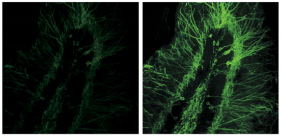
Both maximum intensity projection from 23 slices
Deep Focus Mode improves image brightness

Objectives
- XLPLN10XSVMP, Olympus ultra-long working distance 10X MPE-optimized multi-immersion objective 60NA, WD 8 mm, correct collar for refractive indices 1.33-1.52
- XLPLN25XWMP2, Olympus ultra 25x MPE water-immersion objective 1.05 NA, 2 mm WD
- 5x NA 0.1, WD 20 mm dry (MPLN5X-1-7) and 40x, NA 0.80, WD 3.30 mm water-immersion (LUMPLFLN40XW(F)) objectives on swing nosepiece
Optimized for best live-tissue and intravital multiphoton imaging. Widefield design maximizes fluorescence light capture. Correction collar provides correction for spherical aberration across a wide variety of tissue refractive indices 400nm-1600nm transmission coatings. Chromatically corrected for 700nm to 1300nm IR lasers.
Dichroic(s)
- AOBS serves as tunable dichroic for all lasers

Detectors
- Includes 2 channels of GaAsP PMTs for maximum sensitivity and 2 channels of multi-alkali PMTs for maximum spectral range
- Transmitted light detector for Brightfield/DIC Image Acquisition
Olympus GaAsP detectors provide approximately twice the quantum efficiency of standard multi-alkali PMTs. All non-descanned detectors are positioned as close as possible to the specimen and signaling optics have been enlarged to facilitate maximum detection efficiency of scattered fluorescence.
Computer and Software
- HP Windows 764-bit Workstation and 30” Monitor, 2TB HD, 128 & 256 GB SSDs, 32GB DDR3 RAM
- FVMPE-RS system software including sequence manager, stage control, multipoint mapping, and data analysis
- Full-feature acquisition software provides complete control of system components including laser wavelength and intensity attenuation, laser coupling optics, scanners, & PMTs
- Stage Control Software provides XY motorized stage control, map image acquisition for easy target locating, tiling acquisition, and image stitching
- Multipoint Mapping Software provides high-speed multipoint stimulation, multipoint acquisition, and optogenetic mapping
- Sequence Manager allows programming of complex imaging tasks with microsecond precision within imaging tasks and millisecond precision between imaging tasks for up to two weeks
- Embedded 64-bit 3D rendering tool
- Olympus cellSens Dimension provided for post-acquisition data analysis
Tutorials
Posted under Imaging and Histology


You must be logged in to post a comment.